Microtubules & Kinesin Motors
To construct active materials in the Dogic lab, I borrow bio-materials (proteins and biopolymers) from the biological world which are self-motile, efficient, energy consuming micro-machines. By reconstituting them, we can perform highly tunable experiments in vitro and retain complete experimental control over all parameters in the system.
The Microtubule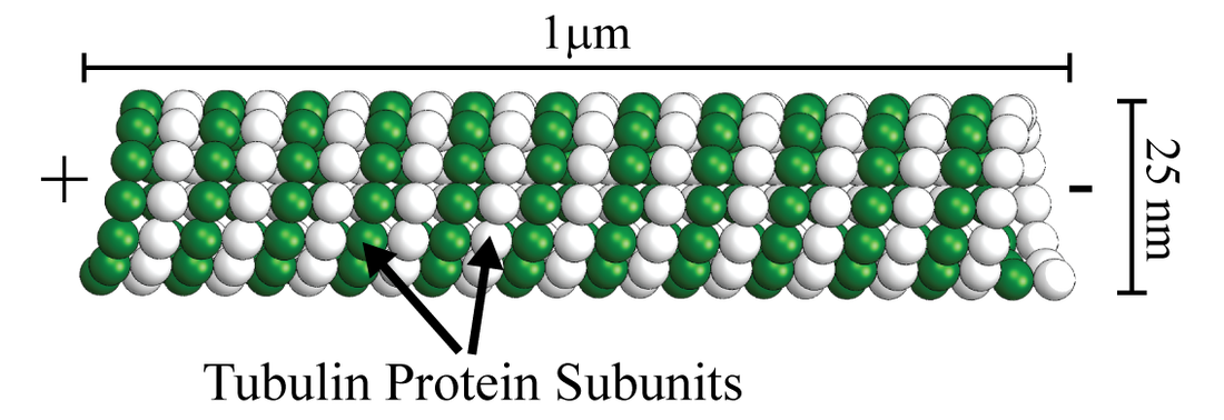
One of the fundamental components in our experiment is a rigid, rod-like filament called a ‘Microtubule’. Microtubules are found throughout all of nature and play a major role in cell division, cellular cytoskeleton structure, and cellular motility. In our model active matter system, the microtubules are about 1 micron in length and are stabilized to prevent dynamic instability from occurring. Individual microtubules are also polar structures. They have a plus end and a minus end.
|
Check out this Youtube video for a great animated visualization of Microtubules and to learn more about their structure and dynamics.
|
Kinesin Motor Clusters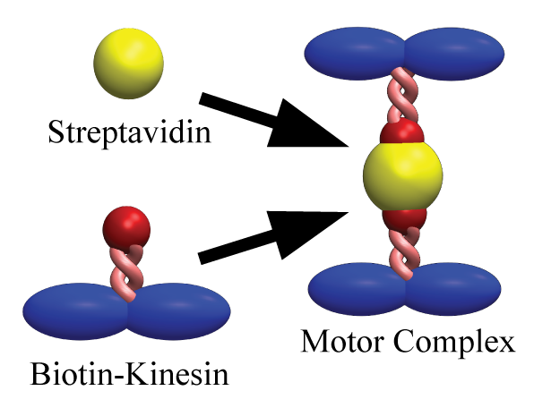
The activity in our system comes from a specialized type of protein which can convert chemical fuel into mechanical energy. The motor protein we use is called Kinesin, and functions by hydrolyzing ATP and undergoing a conformational change which advances the motor along the microtubule substrate, thus effectively allowing the motor to ‘walk’ along a microtubule filament. The Kinesin motor we use preferentially walks towards the plus end of the microtubule filament. To generate activity at the microtubule, we tie multiple kinesin motors together into clusters which are able to crosslink multiple microtubules and exert forces between multiple filaments.
|
This Youtube video explains how a Kinesin motor hydrolyzes ATP in order to move. It is a great animation which depicts the processive walking of a Kinesin motor.
|
Depletion Forms Microtubule Bundles
|
The addition of a non-absorbing polymer, poly-ethylene glycol (PEG), induces an effective attractive interaction between microtubule filaments. This 'bundling' mechanism is entropically driven but can also be thought of in terms of the the PEG exerting an osmotic pressure on the microtubules.
|
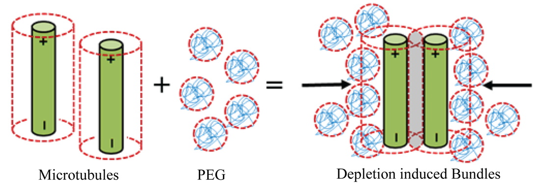
Depletion forces from the polymer PEG induces spontaneous bundling of Microtubule filaments.
|
Extending Microtubule Bundles
|
Combining microtubules, kinesin motor crosslinkers, and depletion agent PEG, results in the formation of microtubule bundles in which the motor clusters drive inter-filament sliding. The inter-filament sliding results in net extension of bundles. We can visualize the extension of microtubule bundles using fluorescence microscopy. In this video, we observe microtubule bundles aligning due to the depletion interaction and then sliding past one another due to motor sliding forces.
|
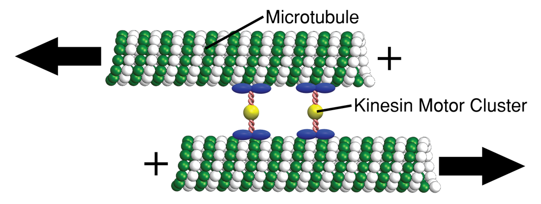
Microtubules slide apart due to forces generated by the Kinesin motor protein clusters.
|
|
|

Microtubules, Kinesin motor clusters, and PEG are the basic active unit in our system.
|
These basic components will act as the building blocks of our model active matter system. The fundamental constituent active particle in our system is an extensile bundle of microtubule filaments. Take a look at how I use these components to study active matter: